Nearly half a year has passed since Heather and I got married in Busan. And if you're not yet married and wondering "So how's married life?" well, it's really similar to life before marriage. Getting married is a good thing overall, but just make sure you choose the right person. Luckily, I did.
Not sure whether she did though...

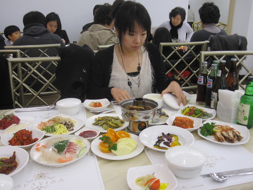
Ben is a Canadian teacher at Heather's new CDI branch and we were invited to the wedding. Heather has only been working a couple of months and I'd never met any of the teachers from the branch before, so it was a nice gesture. The ceremony was held at a wedding hall (I think the name was Suaviss) north of the river.

I heard a one-liner the other day about a woman saying to her best friend "For 18 years my husband and I were the happiest people on Earth. Then we met."
I found it funny, but anyhow I hope Ben and his new wife have a rewarding life together.

If you don't know the bride and groom very well, the best thing about going to a wedding is free food and alcohol. In Korea I guess it's not really free, because all the guests will typically give the couple some money as a gift. A regular amount of money to give would be W50,000 (about US$50). Any more than that is being generous, while anything less is being a bit of a scrooge and only forgivable if you're a grad student.
And yes, they do record your name and how much you gave. Forensic examination of the records afterward constitutes half the fun of getting married.

So let's go back to the lab and see what's going on. These days I'm doing three experiments simultaneously, two of which are not going so well. The one that is working is an ongoing microscopy experiment, which has evolved into using tobacco and rice cells. Earlier this year I managed to get fluorescence in onion cells by microparticle bombardment, but that has since become child's play. I remember the first time I saw fluorescent green in an onion, I literally ran up the stairs to tell Rakshya "I've got signal!"
Now I've seen more than 100 fluorescent images and it's not nearly as exciting.

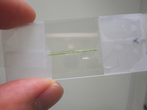
This photo shows my preparation of a rice xylem tube, which can be thought of as a vein that water travels through. Rice is an exceedingly difficult plant to work with. It happens to have every possible element that a plant could have to make it difficult to study. It repels water (and dye), emits light in every wavelength, falls to pieces when finely cut and starts rearranging its cellular 'gizzards' as soon as you put it on a slide. My teaching instincts normally tell me to discipline such an unruly subject, but in this case it would be counter-productive.

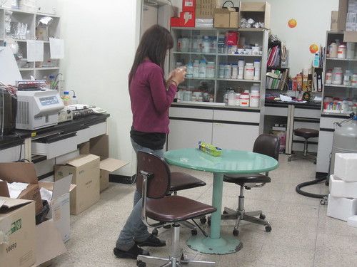
This is Rakshya Singh in her lab at Sejong University. She's a Nepalese PhD student working on Magnaporthe oryzae, a rice fungus. We're starting to collaborate on experiments and it's great to have someone that I can discuss things with.

They also have a confocal microscope, which I use nearly every week. Most Fridays I will pack some plants into a box and travel across town to their university so I can use it. Confocal microscopes are sensitive pieces of equipment and typically cost around US$350,000.
Normal optical microscopes illuminate whatever you're looking at with a backlight, which is fine for simple images, but what you see is a collection of everything that the light passes through. So if you imagine yourself looking straight down at a small opaque man standing on the slide with a hat on, the image you get would show every cross section from his shoes to his hat, all on top of each other. This would make it very difficult to tell what kind of socks he's wearing, for instance. A confocal microscope gets around this by splitting two laser beams and using them as the light source. This allows high precision imaging of any cross section that you want to focus on. You can even take photos of multiple layers in a cell and stitch them together into a 3D image.

This isn't my picture, but it's in the public domain as a patent application so I guess that means I can use it. It shows the basic idea of a confocal microscope, which was invented by Marvin Minsky. Surprisingly, he came up with the idea 30 years too early and it wasn't until lasers became readily available that the concept became feasible.
The straight lines are showing the paths of light being bent and deflected by mirrors and a lens.

And this is the kind of image that you get with a confocal microscope. These are my tobacco cells, after having been attacked with a bacterial strain called Agrobacterium tumefaciens. Before setting the critters loose, I inserted some engineered DNA into them, which causes the cells that they attack to turn bright green.
Tobacco cells are rather oddly shaped, I find.

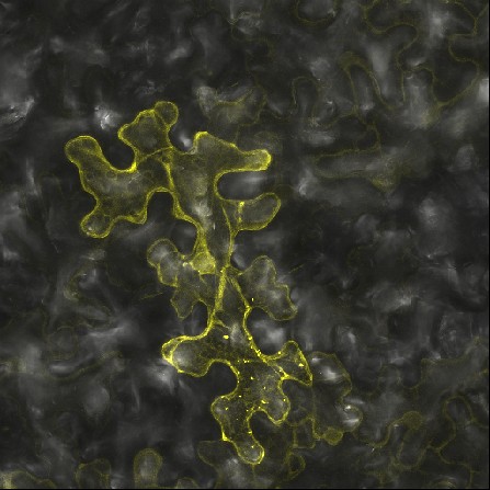
And this is something similar but more interesting. It's a tobacco cell displaying Bi-molecular Fluorescence Complementation. In science terms we would say that it's showing the reconstitution of a fluorophore. In everyday language that means we broke our glowing molecule into two halves, and inserted them separately into the cell. The cell will turn yellow if the two halves find each other. If it doesn't glow it means that they didn't meet up. So this experiment is one of the minority that I'm having success with at the moment. In the bigger picture, this provides evidence that my two proteins are interacting in the plant. One is a bacterial toxin and the other is a plant protein, and I'm trying to figure out what's going on in the grand scheme of things.
Science is all about hits and misses, and keeping good records of both. Hopefully this year will be more fruitful than last.
Not sure whether she did though...

Ben is a Canadian teacher at Heather's new CDI branch and we were invited to the wedding. Heather has only been working a couple of months and I'd never met any of the teachers from the branch before, so it was a nice gesture. The ceremony was held at a wedding hall (I think the name was Suaviss) north of the river.

I heard a one-liner the other day about a woman saying to her best friend "For 18 years my husband and I were the happiest people on Earth. Then we met."
I found it funny, but anyhow I hope Ben and his new wife have a rewarding life together.

If you don't know the bride and groom very well, the best thing about going to a wedding is free food and alcohol. In Korea I guess it's not really free, because all the guests will typically give the couple some money as a gift. A regular amount of money to give would be W50,000 (about US$50). Any more than that is being generous, while anything less is being a bit of a scrooge and only forgivable if you're a grad student.
And yes, they do record your name and how much you gave. Forensic examination of the records afterward constitutes half the fun of getting married.

So let's go back to the lab and see what's going on. These days I'm doing three experiments simultaneously, two of which are not going so well. The one that is working is an ongoing microscopy experiment, which has evolved into using tobacco and rice cells. Earlier this year I managed to get fluorescence in onion cells by microparticle bombardment, but that has since become child's play. I remember the first time I saw fluorescent green in an onion, I literally ran up the stairs to tell Rakshya "I've got signal!"
Now I've seen more than 100 fluorescent images and it's not nearly as exciting.

This photo shows my preparation of a rice xylem tube, which can be thought of as a vein that water travels through. Rice is an exceedingly difficult plant to work with. It happens to have every possible element that a plant could have to make it difficult to study. It repels water (and dye), emits light in every wavelength, falls to pieces when finely cut and starts rearranging its cellular 'gizzards' as soon as you put it on a slide. My teaching instincts normally tell me to discipline such an unruly subject, but in this case it would be counter-productive.

This is Rakshya Singh in her lab at Sejong University. She's a Nepalese PhD student working on Magnaporthe oryzae, a rice fungus. We're starting to collaborate on experiments and it's great to have someone that I can discuss things with.

They also have a confocal microscope, which I use nearly every week. Most Fridays I will pack some plants into a box and travel across town to their university so I can use it. Confocal microscopes are sensitive pieces of equipment and typically cost around US$350,000.
Normal optical microscopes illuminate whatever you're looking at with a backlight, which is fine for simple images, but what you see is a collection of everything that the light passes through. So if you imagine yourself looking straight down at a small opaque man standing on the slide with a hat on, the image you get would show every cross section from his shoes to his hat, all on top of each other. This would make it very difficult to tell what kind of socks he's wearing, for instance. A confocal microscope gets around this by splitting two laser beams and using them as the light source. This allows high precision imaging of any cross section that you want to focus on. You can even take photos of multiple layers in a cell and stitch them together into a 3D image.

This isn't my picture, but it's in the public domain as a patent application so I guess that means I can use it. It shows the basic idea of a confocal microscope, which was invented by Marvin Minsky. Surprisingly, he came up with the idea 30 years too early and it wasn't until lasers became readily available that the concept became feasible.
The straight lines are showing the paths of light being bent and deflected by mirrors and a lens.

And this is the kind of image that you get with a confocal microscope. These are my tobacco cells, after having been attacked with a bacterial strain called Agrobacterium tumefaciens. Before setting the critters loose, I inserted some engineered DNA into them, which causes the cells that they attack to turn bright green.
Tobacco cells are rather oddly shaped, I find.

And this is something similar but more interesting. It's a tobacco cell displaying Bi-molecular Fluorescence Complementation. In science terms we would say that it's showing the reconstitution of a fluorophore. In everyday language that means we broke our glowing molecule into two halves, and inserted them separately into the cell. The cell will turn yellow if the two halves find each other. If it doesn't glow it means that they didn't meet up. So this experiment is one of the minority that I'm having success with at the moment. In the bigger picture, this provides evidence that my two proteins are interacting in the plant. One is a bacterial toxin and the other is a plant protein, and I'm trying to figure out what's going on in the grand scheme of things.
Science is all about hits and misses, and keeping good records of both. Hopefully this year will be more fruitful than last.
1 comment:
nice photos, from both wedding and confocal
Post a Comment